
Nathan Clark Research
I am Nathan Clark, a second year Masters student in the biomedical engineering program here at the University of Rochester. My graduate studies are in the field of biomedical imaging; I research object reconstruction via fluorescence microscopy and phase retrieval under the direction of Professors Richard E. Waugh and James R. Fienup.
Fluorescence microscopy is used in studying many aspects of cellular function including cell interactions, molecular distribution, and dynamics under changing physiological conditions. However, in using conventional fluorescence microscopy, light from out-of-focus portions of the object decreases contrast and does not allow one to correlate image intensity with labeled protein density; multiple focal planes contribute to the overall intensity.
One solution has been the development of confocal microscopy, which blocks light beyond the focal plane using a pinhole aperture. But confocal microscopy is expensive, allows little light through the pinhole, and is not a suitable imaging modality for some samples.
We compute information similar to what one would obtain from confocal microscopy by using information about the microscope, knowledge of the constraints on the object, and nonlinear optimization techniques. Our research includes membrane modeling, microscope characterization, modeling image formation, and synthesizing images, all of which are used to reconstruct the real fluorescence distribution on the surface of a cell.
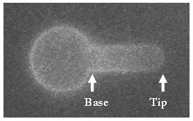
A red blood cell that has been aspirated into a micropipette and imaged using standard fluorescence microscopy. Researchers in cell and membrane mechanics are interested in the protein density at the base of the aspirated cylinder versus the tip. However, at the base of the projection, the intensity could be due to out-of-focus light that originates from the spherical portion of the membrane.
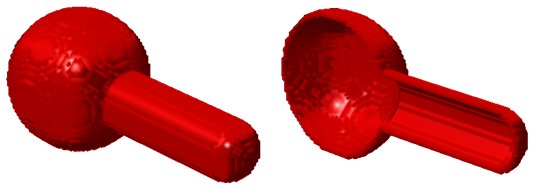
3D rendering (and cut away view) showing our model of the membrane surface area. This model is used to synthesize images taken by the microscope. Before we can synthesize images we must also model our optical system.
We use fluorescent beads to gather an image stack representing the three-dimensional point spread function of the microscope.
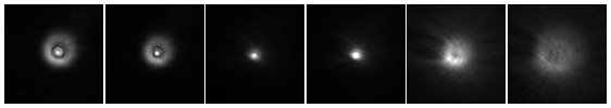
Several images (independently scaled) representing the three-dimensional point spread function of the microscope. These images are used in a phase retrieval algorithm to determine the complex valued exit pupil of the microscope.
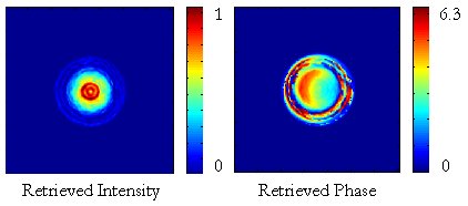
Retrieved intensity and phase of the microscope’s exit pupil.
The retrieved exit pupil and the membrane model are used to synthesize images taken by the microscope.
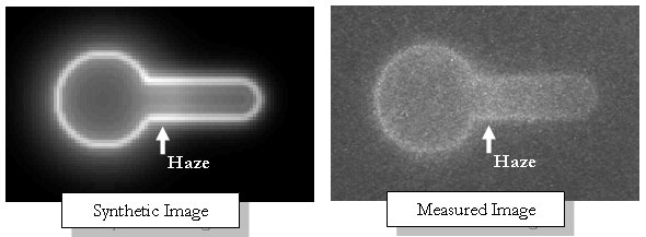
A comparison between the synthetic and measured images shows each has a haze at the base of the aspirated portion of the membrane. This haze can lead to misinterpretation of the density of the labeled protein.
We reconstruct the true fluorophore distribution using measured thru-focus images of the red blood cell, nonlinear optimization, and models of the membrane surface area and the optical system.
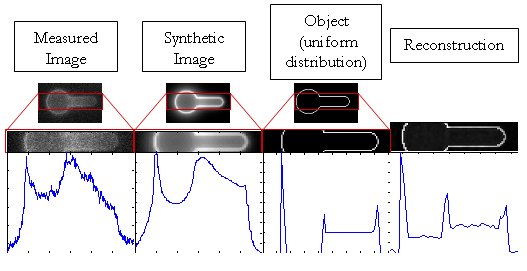
The true fluorophore distribution along the aspirated portion of the membrane is more easily interpreted from the reconstruction than the measured image.